Long Bone Slide Labeled - Seer Training Classification Of Bones - Trabecular bone this slide shows trabecular, or spongy bone.. Free online quiz compact bone microscope slide labeled. A long bone has two main regions: Click on the tags below to find other quizzes on the same subject. The latter is a dense regular collagenous c.t. There is a printable worksheet available for download here so you can take the quiz with pen and paper.
Compact bone diagram labeled 13 photos of the compact bone diagram labeled bone marrow diagram, compact bone diagram quiz, compact bone slide labeled, diagram long bone, labeled compact bone model, human anatomy, bone marrow diagram, compact bone diagram quiz, compact bone slide labeled, diagram long bone, labeled compact bone model This is an online quiz called label a long bone there is a printable worksheet available for download here so you can take the quiz with pen and paper. Рет қаралды 106 м.2 жыл бұрын. A section of monkey lumbar vertebral body showing single (solid arrow) and. A long bone has two main regions:
Chapter 6 Page 7 Histologyolm from stevegallik.org Bone tissue is the structural component of bones. Compact bone diagram labeled 13 photos of the compact bone diagram labeled bone marrow diagram, compact bone diagram quiz, compact bone slide labeled, diagram long bone, labeled compact bone model, human anatomy, bone marrow diagram, compact bone diagram quiz, compact bone slide labeled, diagram long bone, labeled compact bone model Long bones have a thick outside layer of compact bone and an inner medullary cavity containing bone marrow. 450 x 450 jpeg 54 кб.this is the diagram of label bone diagram that you search. Slide 31 hyaline cartilage (lavender matrix), with perichondrium (pink) outside it. Click on the tags below to find other quizzes on the same subject. The surface of trabeculae are covered by a thin layer of inactive cells called endosteum. Labeled compact bone microscope slides | labeled histology slides.
This is an online quiz called label a long bone there is a printable worksheet available for download here so you can take the quiz with pen and paper.
They have a shaft part that connects the two ends referred to as epiphysis (mostly spongy bone with a thin layer of compact bone). The latter is a dense regular collagenous c.t. The surface of trabeculae are covered by a thin layer of inactive cells called endosteum. The epiphyses are at the ends of the long bone and are the parts of the bone that participate in joint surfaces. Long bone model labeled / front. Labeled compact bone microscope slides | labeled histology slides. Calcein labels in cancellous bone fluorescent microscopy calcein labels (magnification: The diaphysis is the hollow, tubular shaft that runs between the proximal and distal ends of the bone. 40x (spongy bone, osteoblasts, osteoclasts, appositional bone growth on surface of long bone). Such a change has begun in the middle of the shaft of this bone, thanks to the invasion of blood vessels and their accompanying Cavity within the shaft of the long bones filled with bone marrow. Skeleton system histology slides med lab microscope slides phlebotomy college hacks anatomy and physiology love my job biology. Hi all, i have uploaded a new medical animation tutorial.
The ends of a long bone contain spongy bone and an epiphyseal line. Human blood smear, giemsa stain, 86x scan from hematopathology normals collection (this slide contains 3 basophil cells) virtual slide. Label a long bone shannan muskopf october 16, 2020 anatomy students in traditional classes may do practice labeling the bone on paper or even doing a coloring activity to help them learn the parts of the bone. The diaphysis is the hollow, tubular shaft that runs between the proximal and distal ends of the bone. Compact bone diagram labeled 13 photos of the compact bone diagram labeled bone marrow diagram, compact bone diagram quiz, compact bone slide labeled, diagram long bone, labeled compact bone model, human anatomy, bone marrow diagram, compact bone diagram quiz, compact bone slide labeled, diagram long bone, labeled compact bone model
Spongy Cancellous Bone Slide Diagram Quizlet from o.quizlet.com Label a long bone shannan muskopf october 16, 2020 anatomy students in traditional classes may do practice labeling the bone on paper or even doing a coloring activity to help them learn the parts of the bone. Dense irregular connective tissue that lines the medullary cavities of long bones. Slide 31 hyaline cartilage (lavender matrix), with perichondrium (pink) outside it. Рет қаралды 106 м.2 жыл бұрын. Spongy bone shown from slide of rib, includes endosteum and marrow space along with osteoblasts lining the bone and periosteum on the outside. Slide 61 all of the long bones and many others of the body, are preformed embryologically in hyaline cartilage and then replaced by bone by endochondral ossification. Two types of bone tissues in cross section of a long bone : Ground bone cancellous bone demineralized bone trabecular bone central canal bone lamellar spongy bone lacuna bone cortical bone decalcified tooth decalcified enamel decalcified heart.
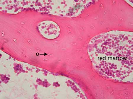
Same bone section (slide 24) at higher magnification.
The surface of trabeculae are covered by a thin layer of inactive cells called endosteum. Compact bone diagram labeled 13 photos of the compact bone diagram labeled bone marrow diagram, compact bone diagram quiz, compact bone slide labeled, diagram long bone, labeled compact bone model, human anatomy, bone marrow diagram, compact bone diagram quiz, compact bone slide labeled, diagram long bone, labeled compact bone model Free online quiz compact bone microscope slide labeled. This is an online quiz called long bone anatomy. Labeled compact bone microscope slides | labeled histology slides. Trabecular bone this slide shows trabecular, or spongy bone. Inside the diaphysis is the medullary cavity, which is filled with yellow bone marrow in an adult. Try pressing the section on the slide to ensure that the layer of glue is as thin as possible. Click on the tags below to find other quizzes on the same subject. The latter is a dense regular collagenous c.t. Skeleton system histology slides med lab microscope slides phlebotomy college hacks anatomy and physiology love my job biology. The epiphyses are at the ends of the long bone and are the parts of the bone that participate in joint surfaces. Fetal leg, cross section, masson.
They have a shaft part that connects the two ends referred to as epiphysis (mostly spongy bone with a thin layer of compact bone). Attach the ground side to the slide using. A long bone has two main regions: Skeletal system of a frog frog bone. This slide shows endochondral ossification, the process by which cartilage is calcified to form bone.
Spongy Bone from eugraph.com 450 x 450 jpeg 54 кб.this is the diagram of label bone diagram that you search. Dense irregular connective tissue that lines the medullary cavities of long bones. Two types of bone tissues in cross section of a long bone : Ground bone cancellous bone demineralized bone trabecular bone central canal bone lamellar spongy bone lacuna bone cortical bone decalcified tooth decalcified enamel decalcified heart. Human blood smear, giemsa stain, 86x scan from hematopathology normals collection (this slide contains 3 basophil cells) virtual slide. Labeled compact bone microscope slides | labeled histology slides. Identify portions of three long bones, arranged in a linear fashion, separated from each other by a relatively narrow clear space (the joint space) and surrounded by soft tissues. The diaphysis and the epiphysis (figure 6.3.1).
Saved by university of colorado boulder.
This labeling is simply a drag and drop exercise that students can complete directly in google slides. A flat bone is characterized by parallel surfaces of figure figure observe a slide preparation labeled ground bone; Some, mostly older, compact bone is remodelled to form these haversian systems (or osteons).the osteocytes sit in their lacunae in concentric rings around a central haversian canal (which runs longitudinally).the osteocytes are arranged in concentric rings of bone matrix called lamellae (little plates), and their processes run in interconnecting canaliculi. Spongy bone shown from slide of rib, includes endosteum and marrow space along with osteoblasts lining the bone and periosteum on the outside. This slide shows endochondral ossification, the process by which cartilage is calcified to form bone. Slide 61 all of the long bones and many others of the body, are preformed embryologically in hyaline cartilage and then replaced by bone by endochondral ossification. Compact bone diagram labeled 13 photos of the compact bone diagram labeled bone marrow diagram, compact bone diagram quiz, compact bone slide labeled, diagram long bone, labeled compact bone model, human anatomy, bone marrow diagram, compact bone diagram quiz, compact bone slide labeled, diagram long bone, labeled compact bone model Long bone model labeled / front. Two types of bone tissues in cross section of a long bone : Inside the diaphysis is the medullary cavity, which is filled with yellow bone marrow in an adult. Skeletal system of a frog frog bone. Free online quiz compact bone microscope slide labeled. Bone models with a thin cortical layer and an open cell.
A flat bone is characterized by parallel surfaces of figure figure observe a slide preparation labeled ground bone; long bone labeled. A long bone has two main regions:
Berbagi
Posting Komentar
untuk "Long Bone Slide Labeled - Seer Training Classification Of Bones - Trabecular bone this slide shows trabecular, or spongy bone."


Posting Komentar untuk "Long Bone Slide Labeled - Seer Training Classification Of Bones - Trabecular bone this slide shows trabecular, or spongy bone."